

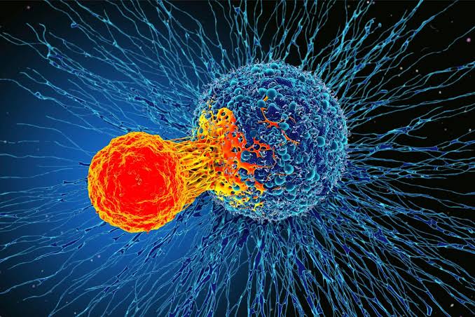
A new study has discovered that nearby conditions and nutrients influence how cancer cells evade immune cells and how that protection might be disrupted. Cancer cells can sometimes escape immune attack by covering themselves in a dense layer of sugar-derived molecules that makes them harder for immune cells to recognise.
A research from Sanford Burnham Prebys Medical Discovery Institute and collaborators across North America now suggests that this protective coating is shaped not just by the cancer cell itself, but also by the physical and nutritional conditions surrounding the tumor.
The findings, published in Science Advances, show how the tumor microenvironment, which includes nearby immune cells, connective tissue, blood vessels, proteins, and carbohydrates, can influence this sugar-rich barrier. The researchers also identified a potential way to reduce the coating so immune cells can once again recognise and eliminate cancer cells.
Lead and corresponding author of the study, Dr. Kevin Tharp had previously observed that physical pressure on cells can alter how their mitochondria function. Because solid tumors often subject cells to unusually stiff surroundings, he suspected those physical conditions might help explain some of the metabolic changes commonly seen in cancer. “Primary tumors are typically stiffer than their surrounding tissue. “This led me to hypothesise that the biophysical properties of cells influence the altered metabolic programs that everyone observes in tumors,” said Tharp, an assistant professor in the Cancer Metabolism and Microenvironment Program at the Sanford Burnham Prebys NCI-Designated Cancer Centre.
One prominent metabolic feature of tumors is reduced oxidative metabolism of glucose, the process cells use to extract energy from glucose with the help of oxygen. Previous research has shown that this shift can depend on which nutrients are available around the cells rather than being an unavoidable property of cancer cells themselves.
To separate these influences, Tharp and his colleagues grew cells under several combinations of physical and nutritional conditions. Some cells were placed in stiff environments resembling those around primary tumors, while others experienced softer conditions closer to healthy tissue. The researchers also compared a standard laboratory culture medium with a newer medium designed to more closely reproduce the nutrient composition found in the human body.
Both were tested under normal and elevated glucose conditions to model hyperglycemia. Changing these conditions altered which proteins the cells produced, the concentrations of metabolites inside them, and the thickness of the sugarderived surface layer known as the glycocalyx.
Crucially, excess glucose increased the depth of this protective coating only when cells were grown in the more physiologically realistic medium. “We observed that changing the physiological media composition and changing the available metabolites for those tumor cells reveals distinct biology for normal and tumor cell metabolism,” said Tharp.
The researchers next followed the metabolic changes into the molecular building blocks of the glycocalyx. This outer coating is composed of glycoconjugates, molecules in which carbohydrates are attached to proteins or lipids. Because glucose can supply material for constructing glycoconjugates, changes in glucose availability offered a possible explanation for why the coating became thicker.